The Bloodpressure Program™ By Christian Goodman The procedure is a very basic yet effective method to lessen the effects of high blood pressure. To some people, it sounds insane that just three workouts in a day can boost fitness levels and reduce blood pressure simultaneously. The knowledge and research gained in this blood pressure program were really impressive.
Eye Damage Related to Hypertension
Hypertension, or high blood pressure, is a condition that affects multiple organs and systems in the body, including the eyes. Chronic high blood pressure can lead to a variety of eye-related complications, collectively known as hypertensive retinopathy, as well as other conditions like choroidopathy and optic neuropathy. These conditions can cause significant visual impairment and even blindness if left untreated. This comprehensive guide explores the relationship between hypertension and eye damage, the mechanisms involved, the symptoms and stages of hypertensive eye disease, and strategies for prevention and management.
The Relationship Between Hypertension and Eye Health
The eyes are highly vascular organs, meaning they contain many blood vessels, particularly in the retina, the light-sensitive layer at the back of the eye. The retina is crucial for vision, as it converts light into neural signals that are sent to the brain. High blood pressure can cause damage to these delicate blood vessels, leading to a range of eye conditions.
1. Hypertensive Retinopathy
Hypertensive retinopathy is the most common form of eye damage related to hypertension. It occurs when high blood pressure causes changes to the retinal blood vessels, leading to impaired vision.
- Mechanism: Chronic high blood pressure exerts increased force against the walls of the retinal blood vessels, causing them to thicken, narrow, or become damaged. This can reduce blood flow to the retina and lead to a variety of complications, including retinal hemorrhages (bleeding), exudates (fluid leakage), and edema (swelling).
- Consequences: If left untreated, hypertensive retinopathy can lead to progressive vision loss and even blindness. The severity of retinopathy often correlates with the duration and severity of hypertension.
2. Hypertensive Choroidopathy
Hypertensive choroidopathy is a less common condition that affects the choroid, a layer of blood vessels that supplies oxygen and nutrients to the outer layers of the retina.
- Mechanism: High blood pressure can cause changes in the choroidal blood vessels, leading to ischemia (reduced blood flow) and fluid accumulation under the retina (serous retinal detachment). This condition is more likely to occur in younger individuals with acute, severe hypertension, such as in malignant hypertension or preeclampsia during pregnancy.
- Consequences: Hypertensive choroidopathy can cause visual disturbances, such as blurry vision or blind spots, and can lead to more serious complications if not promptly treated.
3. Hypertensive Optic Neuropathy
Hypertensive optic neuropathy is a condition in which high blood pressure damages the optic nerve, the nerve responsible for transmitting visual information from the eye to the brain.
- Mechanism: Elevated blood pressure can reduce blood flow to the optic nerve, leading to ischemia and subsequent damage. This condition is often associated with malignant hypertension, where blood pressure rises rapidly and to very high levels.
- Consequences: Optic neuropathy can result in vision loss, ranging from partial loss of peripheral vision (in the early stages) to complete blindness if the entire optic nerve is affected.
Stages and Symptoms of Hypertensive Eye Disease
Hypertensive eye disease progresses through several stages, each with its own set of symptoms and clinical signs. The severity of the eye damage typically corresponds to the severity and duration of hypertension.
1. Mild Hypertensive Retinopathy
- Description: In the early stages of hypertensive retinopathy, changes to the retinal blood vessels are minimal and may not cause noticeable symptoms. This stage is often detected during routine eye examinations.
- Symptoms: Most individuals with mild hypertensive retinopathy do not experience symptoms. However, some may notice subtle changes in vision, such as mild blurring or difficulty seeing in low light.
- Clinical Signs: Ophthalmologists may observe narrowing of the retinal arteries (arteriolar narrowing) and an increase in the light reflex along the arteries (copper wiring) during an eye exam.
2. Moderate Hypertensive Retinopathy
- Description: As hypertensive retinopathy progresses, the damage to the retinal blood vessels becomes more pronounced, leading to more noticeable symptoms and signs.
- Symptoms: Individuals may experience more significant visual disturbances, such as blurred vision, dark spots, or flashes of light.
- Clinical Signs: Ophthalmologists may observe more severe changes, including arteriovenous (AV) nicking, where the retinal veins are compressed by the overlying arteries, as well as retinal hemorrhages, cotton wool spots (areas of retinal ischemia), and hard exudates (lipid deposits).
3. Severe Hypertensive Retinopathy
- Description: In severe hypertensive retinopathy, the retinal blood vessels are severely damaged, leading to significant visual impairment.
- Symptoms: Symptoms may include significant vision loss, blind spots, or a sudden loss of vision if the condition is associated with a retinal vein occlusion or retinal detachment.
- Clinical Signs: Severe hypertensive retinopathy is characterized by extensive retinal hemorrhages, cotton wool spots, exudates, and retinal edema. Papilledema (swelling of the optic disc) may also be present, indicating a more serious and potentially life-threatening condition such as malignant hypertension.
4. Hypertensive Choroidopathy and Optic Neuropathy
- Hypertensive Choroidopathy:
- Symptoms: Visual symptoms may include blurry vision, distorted vision (metamorphopsia), or blind spots (scotomas). These symptoms are often associated with acute, severe hypertension.
- Clinical Signs: Ophthalmologists may observe serous retinal detachment (fluid accumulation under the retina) and Elschnig spots (areas of choroidal infarction).
- Hypertensive Optic Neuropathy:
- Symptoms: Symptoms of optic neuropathy include loss of peripheral vision, decreased visual acuity, and, in severe cases, complete vision loss.
- Clinical Signs: Ophthalmologists may observe optic disc swelling, pallor, and signs of ischemic optic neuropathy during an eye exam.
Risk Factors for Hypertensive Eye Disease
Several factors increase the risk of developing hypertensive eye disease:
- Duration and Severity of Hypertension: The longer a person has uncontrolled high blood pressure, and the higher their blood pressure levels, the greater the risk of developing hypertensive eye disease.
- Age: Hypertensive retinopathy is more common in older adults, as the risk of hypertension increases with age.
- Diabetes: Diabetes is a significant risk factor for both hypertension and eye disease, and the combination of these conditions can exacerbate the severity of hypertensive retinopathy.
- Smoking: Smoking can damage blood vessels and exacerbate the effects of hypertension on the eyes, increasing the risk of hypertensive eye disease.
- Pregnancy: Conditions such as preeclampsia and eclampsia, which involve high blood pressure during pregnancy, can lead to acute and severe hypertensive eye disease.
- Kidney Disease: Chronic kidney disease, often associated with hypertension, can increase the risk of developing hypertensive retinopathy and other eye conditions.
Prevention and Management of Hypertensive Eye Disease
Preventing and managing hypertensive eye disease involves controlling blood pressure, monitoring eye health, and, when necessary, seeking prompt treatment for any eye-related complications.
1. Blood Pressure Control
The most effective way to prevent hypertensive eye disease is to manage blood pressure through lifestyle modifications and, if necessary, medication. Key strategies include:
- Dietary Approaches: The DASH (Dietary Approaches to Stop Hypertension) diet is particularly effective for managing blood pressure. This diet emphasizes fruits, vegetables, whole grains, lean proteins, and low-fat dairy while reducing sodium, saturated fats, and added sugars.
- Sodium Reduction: Reducing sodium intake to less than 2,300 mg per day, or ideally less than 1,500 mg per day, can lower blood pressure and reduce the risk of hypertensive eye disease.
- Weight Management: Maintaining a healthy weight through a balanced diet and regular physical activity is crucial for controlling blood pressure. Even modest weight loss can lead to significant reductions in blood pressure.
- Regular Physical Activity: Engaging in regular aerobic exercise, such as walking, swimming, or cycling, for at least 150 minutes per week, can help lower blood pressure and protect against hypertensive eye disease.
- Smoking Cessation: Quitting smoking can lead to immediate and long-term improvements in blood pressure and vascular health, reducing the risk of hypertensive eye disease.
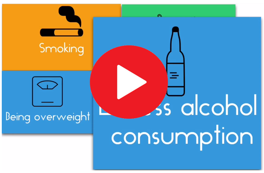
- Alcohol Moderation: Limiting alcohol intake to no more than one drink per day for women and two drinks per day for men can help control blood pressure and reduce the risk of hypertensive eye disease.
- Stress Management: Chronic stress can contribute to elevated blood pressure. Stress-reducing techniques, such as meditation, deep breathing exercises, and yoga, can help lower blood pressure and protect eye health.
2. Regular Eye Examinations
Routine eye examinations are crucial for detecting early signs of hypertensive eye disease and preventing its progression. Individuals with hypertension should undergo regular eye exams, which may include:
- Fundoscopy: A fundoscopy is an examination of the retina and optic nerve using an ophthalmoscope. This test allows the ophthalmologist to detect signs of hypertensive retinopathy, such as arteriolar narrowing, AV nicking, hemorrhages, exudates, and papilledema.
- Optical Coherence Tomography (OCT): OCT is a non-invasive imaging test that provides detailed cross-sectional images of the retina, allowing the detection of retinal edema and other structural changes.
- Fluorescein Angiography: This test involves injecting a fluorescent dye into a vein and taking photographs of the retina as the dye passes through the blood vessels. It helps detect areas of retinal ischemia, leakage, and other abnormalities.
3. Pharmacological Management
For individuals with hypertension and hypertensive eye disease, antihypertensive medications are essential for controlling blood pressure and preventing further eye damage. The choice of medication depends on the individual’s overall health, the severity of hypertension, and the presence of comorbidities.
- Common Antihypertensive Medications:
- ACE Inhibitors and ARBs: These medications are particularly effective for protecting blood vessels, including those in the eyes, from damage by lowering blood pressure and reducing proteinuria.
- Calcium Channel Blockers: These medications relax blood vessels and improve blood flow, helping to lower blood pressure and protect against hypertensive eye disease.
- Diuretics: Diuretics help reduce blood volume by promoting the excretion of sodium and water, thereby lowering blood pressure and reducing the risk of hypertensive eye disease.
- Beta-Blockers: Beta-blockers reduce heart rate and the force of heart contractions, lowering blood pressure and protecting against hypertensive eye disease.
4. Treatment of Eye Complications
If hypertensive eye disease has progressed to a more severe stage, additional treatments may be necessary to manage complications and preserve vision:
- Laser Therapy: In cases of retinal hemorrhages or retinal vein occlusion, laser therapy may be used to seal leaking blood vessels and prevent further damage.
- Anti-VEGF Injections: For individuals with retinal edema or neovascularization (the growth of abnormal blood vessels), anti-VEGF (vascular endothelial growth factor) injections may be administered to reduce swelling and prevent the growth of abnormal vessels.
- Surgery: In severe cases, such as retinal detachment or advanced optic neuropathy, surgical interventions may be required to preserve vision.
Conclusion
Hypertension is a major risk factor for eye damage, including conditions such as hypertensive retinopathy, choroidopathy, and optic neuropathy. These conditions can lead to significant visual impairment and even blindness if left untreated. The relationship between hypertension and eye health underscores the importance of controlling blood pressure through lifestyle modifications, medication, and regular monitoring. By taking proactive steps to manage hypertension and undergoing routine eye examinations, individuals can protect their vision and reduce the risk of hypertensive eye disease. Early detection and prompt treatment are key to preserving vision and maintaining overall eye health in individuals with hypertension.

The Bloodpressure Program™ By Christian Goodman The procedure is a very basic yet effective method to lessen the effects of high blood pressure. To some people, it sounds insane that just three workouts in a day can boost fitness levels and reduce blood pressure simultaneously. The knowledge and research gained in this blood pressure program were really impressive.
